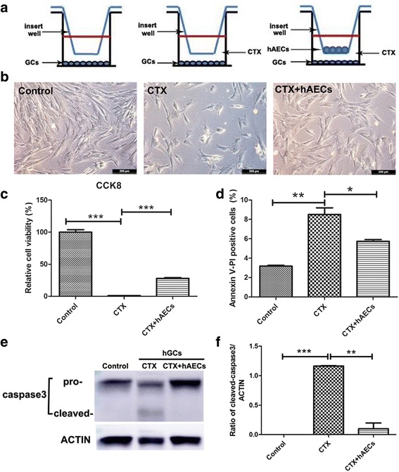
Fig. 4.

Effect of hAECs-secreting cytokines on CTX-induced apoptosis within human granulosa-lutein (hGL) cells in vitro. a 1 × 10⁵ hAECs were seeded on the upper co-culture inserts and 1 × 10⁵ hGL cells were seeded at the bottom of a six-well culture plate with or without CTX. b Bright field image of hGL cells on the bottom of co-culture system in different groups. c and d CCK-8 assay and flow cytometry were used to detect cell viability and apoptosis, respectively. e and f Western blot was performed to evaluate the level of caspase3 and cleaved caspase3 protein expression in hGL cells. Amount of protein loaded was normalized against actin. Data represent means ± SEM. Control group, n = 4; CTX group, n = 4; CTX + hAECs group, n = 4. * P < 0.05; ** P < 0.01; *** P < 0.001. Scale bar is 200 μm in B
