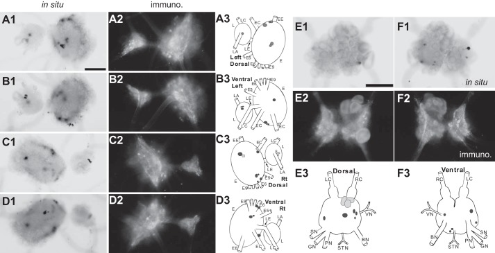
Figure 5.
Distribution of ALK-positive neurons and fibers in the pleural and pedal ganglia and the abdominal ganglion (whole mounts). A–D, left dorsal (A), left ventral (B), right dorsal (C), and right ventral (D) pedal ganglia. E and F, dorsal (E) and ventral (F) abdominal ganglia. 1, in situ hybridization (in situ); 2, immunohistochemistry (immuno.); 3, composite drawings of ALK neurons. Darker shades of gray indicate more intense staining. Scale bar (in A1 and E1), 500 μm. Pleural-pedal abbreviations are as follows. Rt, right; L, pleural ganglion; E, pedal ganglion; LE, pleuropedal connective; EE, pedal commissure; EC, cerebropedal connective; LC, cerebropleural connective; LA, pleuroabdominal connective; E5, posterior tegumentary nerve (P5); E6, anterior parapodial nerve (P6); E9, posterior pedal nerve (P9). Abdominal abbreviations are as follows. LC, left pleuroabdominal connective; RC, right pleuroabdominal connective; VN, vulvar nerve; BN, branchial nerve; STN, spermathecal nerve; PN, pericardial nerve; GN, genital nerve; SN, siphon nerve. For simplicity, not all nerves in the pleural and pedal ganglia were drawn.

