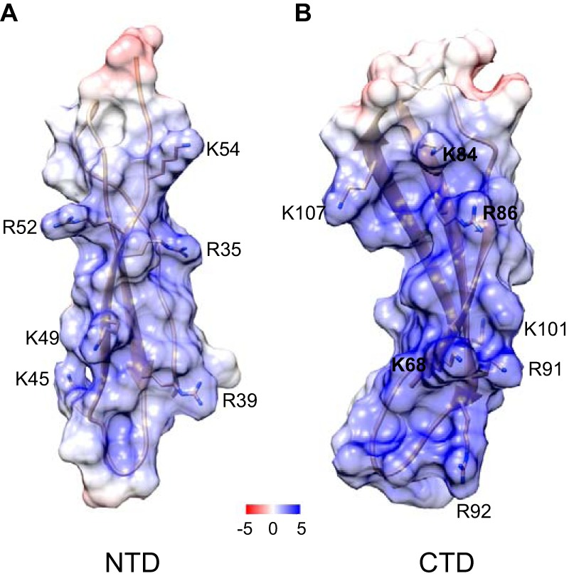
Figure 9.

Location of the αMI domain-binding residues in NTD and CTD of PTN. The electrostatic potential mapped is onto the ribbon diagram of NTD (A) and CTD (B). The unit of potential is in kT·e−1. Positively charged residues identified in the αMI domain binders using peptide libraries are shown. The PTN models are based on Protein Data Bank code 2N6F.
