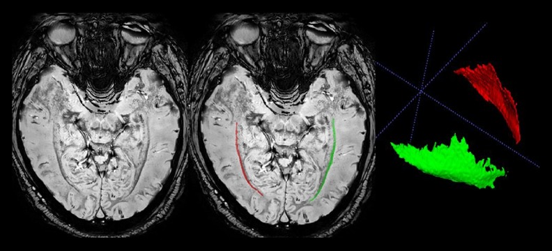
Figure 1.
Representative axial sections of phase difference enhanced (PADRE) images. The characteristic 4 layers, presumably corresponding to the tapetum (a high-signal intensity layer), internal sagittal stratum (a median-signal intensity layer), external sagittal stratum (a low-signal intensity layer), and adjacent white matter (a high-signal intensity layer), are visible beside the lateral ventricles. The external sagittal stratum can be clearly noted as the band with the lowest signal intensity, which differentiates it from surrounding tissue. The red and green regions of interest demonstrate the right and left external sagittal stratum, respectively.

