Fig. 7.

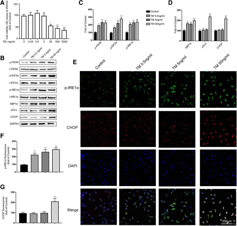
Low doses of TM activate a nontoxic, mild UPR in primary cultured microglia. a Primary microglia were treated with TM (0.02 to 2000 ng/ml) for 24 h followed by assessment of cell viability using the CCK-8 assay. b The expression levels of p-PERK, p-EIF2α, p-IRE1α, XBP1s, ATF4, and CHOP in primary microglia were detected by Western blotting using specific antibodies. Each blot is representative of three experiments. c Phosphorylated levels of PERK, EIF2α, and IRE1α were quantified and normalized to corresponding total levels. d The expression levels of XBP1s, ATF4, and CHOP were quantified and normalized to GAPDH levels. e Cells were stained with p-IRE1α and CHOP antibodies. p-IRE1α-immunopositive (green) and CHOP-immunopositive (red) expression in primary microglia was observed using confocal scanning. Blue staining represents DAPI. Scale bar, 200 μm. f, g Quantitative data of the mean intensity of p-IRE1α and CHOP fluorescence in primary microglia. Each value was then expressed relative to that of the naïve group, which was set to 100. All experiments were repeated three times. *P < 0.05, **P < 0.01 vs. naïve group. The data are presented as the mean ± SEM
