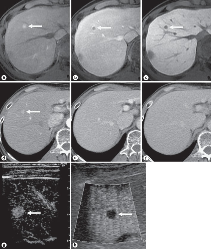
Fig. 1.
Images in a 55-year-old man with chronic hepatitis B developing recurrence of hypervascular HCC 9 months after curative treatment of HCC with RFA. The tumor (arrow) in segment 8, 0.8 cm in diameter, is hyperintense on the arterial phase (a) and hypointense on the portal venous phase (b) and hepatobiliary phase (c) of Gd-EOB-DTPA-enhanced MRI. The corresponding multiphasic CT image obtained during the arterial phase (d) shows a hypervascular nodule, but the nodule shows isoattenuation on the portal venous (e) and equilibrium (f) phase images. Contrast-enhanced ultrasonography with Sonazoid® shows a hypervascular nodule on the early vascular phase image (g) and a clear perfusion defect on the postvascular phase image (h).

