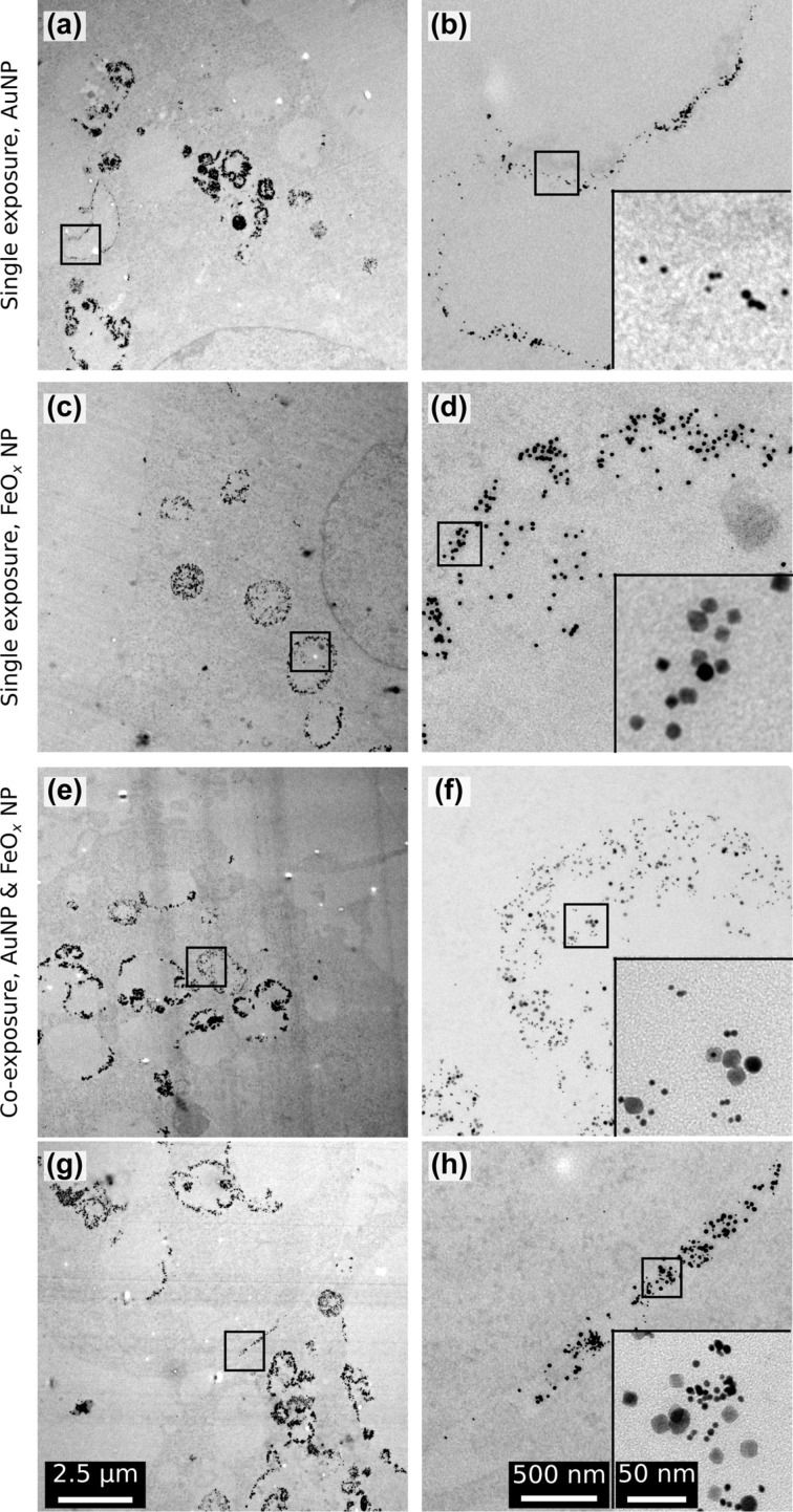
Figure 4.
The intracellular localisation of the NPs 24 h after exposure. (a) Overview showing a part of a cell after exposure only to AuNPs with a number of AuNP-containing vesicles. (b) The higher magnification of the square in (a) reveals that particles are inside the vesicle and appearing both as single entities and aggregates (inset). (c) Overview showing a part of a cell after exposure to only FeOxNPs with FeOxNPs-filled vesicles. (d) The particles appear loosely aggregated (inset) and are contained within the border of the vesicle. (e) Overview showing a part of a cell after co-exposure with AuNPs and FeOxNPs. (f) Both particle types co-appear inside vesicles, either as single particles or small aggregates (inset). (g) A small extracellular crevasse between two cells co-exposed with AuNPs and FeOxNPs is filled with both particle types. (h) Both particle types are mixed extracellularly, in single or aggregated status (inset). All overviews, higher magnifications and insets have a scale bar of 2.5µm, 500 nm and 50 nm, respectively.

