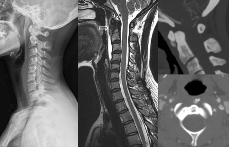
Figure 2.

The x-ray revealed diffuse swelling on the retropharyngeal space. Neck CT showed calcific deposit on C1-2 joint. MRI showed a nodular lesion which located the insertion site of superior oblique tendon of longus colli (white arrow) and retropharyngeal fluid.
