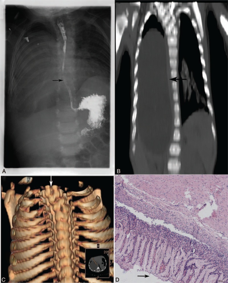
Figure 1.

Imaging findings of a case with esophageal duplication cyst with asymptomatic hemivertebrae. In this 6-month-old male infant, barium swallow examination was inconclusive (A). Computed tomography scan showed an esophageal duplication cyst (arrow), whose coronal is depicted. The cyst was 11.9 × 5.4 × 5.1 cm, with an average wall thickness of 0.5 cm, compressing the trachea, right main bronchus, right inferior lobe (anteriorly), and liver (B). Three-dimensional computed tomography reconstruction showed hemivertebrae located in the upper thoracic spine (T4 and T3) (C). Histopathological examination after hematoxylin and eosin staining showed gastrointestinal-type mucosa (magnification, ×40) (D).
