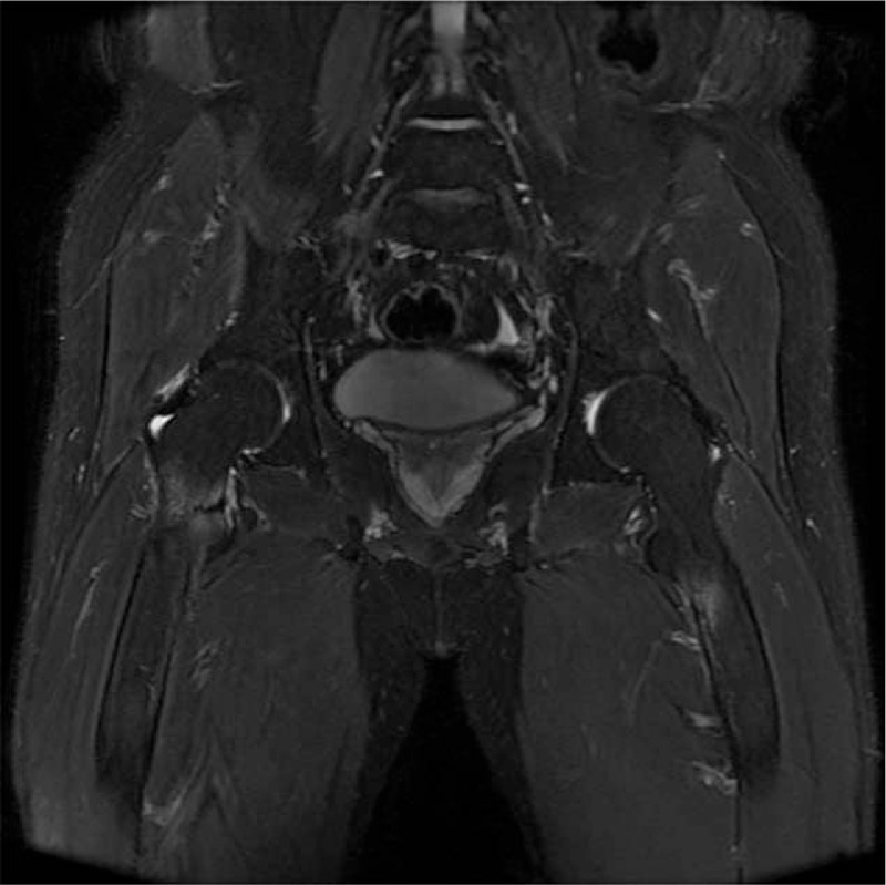
Figure 1.

Fat-saturated coronal magnetic resonance (MR) image revealed hypointensity lesions and cortical bone destruction at the iliopsoas muscle attachment site of the lesser trochanter of right femur. Similar lesion can observe in the lesser trochanter of left femur.
