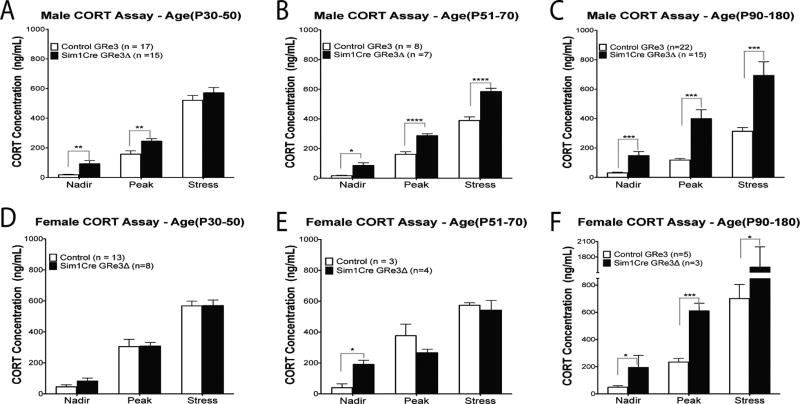
Figure 2. Plasma corticosterone concentrations measured at circadian nadir and peak, and after 20-minutes of restraint stress in males (A, B, C) and female mice (D, E, F).
(A) Male Sim1Cre-GRe3Δ mice at ages P30–P50 (P: postnatal day; n=15) display increased plasma corticosterone at nadir and peak and but not after stress compared to control mice (n=17). (B) Sim1Cre-GRe3Δ male mice at ages P51–P70 (n=7) display increased plasma corticosterone at nadir and peak, as well as after stress compared to control mice (n=8). (C) Adult Sim1Cre-GRe3Δ male mice (n=15) at ages P90–180 compared to control mice (n=22) have elevated corticosterone concentration at nadir, peak and after stress. Circadian rhythm of corticosterone secretion was maintained in males, regardless of age and genotype. (D) Between age P30–P50, corticosterone concentrations of female Sim1Cre-GRe3Δ mice (n=8) did not differ from control mice (n=13) and circadian rhythm was maintain in both genotypes. (E) Female Sim1Cre-GRe3Δ mice ages P51–P70 (n=4) display increased plasma corticosterone at nadir, but no differences in peak and stress –induced corticosterone compared to control mice (n=3). Circadian rhythm of corticosterone secretion was less in female Sim1Cre-GRe3Δ mice at ages P51–P70. (F) Adult Sim1Cre-GRe3Δ female mice (n=3) at ages P90–180 have elevated corticosterone concentration at nadir, peak and after stress compared to controls (n=5). Adult data in C and F are adapted with permission from previously published results (Fig 3, Laryea et al. 2013). *P < 0.05, **P < 0.01, ****P < 0.0001, two-way ANOVA with Bonferonni post-hoc test. Data are shown as mean +/− s.e.m. Abbrev.: CORT-corticosterone.

