Figure 1.
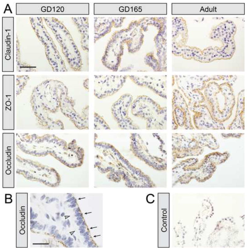
A) Immunoreactivity for claudin-1, ZO-1 and occludin in choroid plexus of gestational day (GD) 90, GD165 fetuses and in adults. Immunoreactivity in tissue was only visible at the apical cell membrane of the epithelial cells and not in other parts of the plexus. There was no apparent difference in the staining pattern in fetuses compared to adult tissue. Tissue was counterstained with hematoxylin. B) Higher power image of occludin staining showing that staining was present on the apical side of epithelial cells (arrows), where the tight-junctions are situated, whereas no staining was present on the basal side of cells (arrowheads) in fetuses. C) Control slides where primary antibodies were omitted resulted in no staining of plexus. Scale bar is 50μm in A and 20μm in C.

