
Fig. 3. BET inhibition reduces homologous recombination.
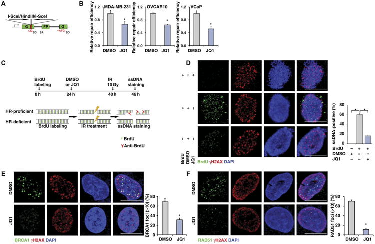
(A) Schematic illustration of HR reporter assay. SD, splice donor; SA, splice acceptor; G, green; FP, fluorescent protein; ATG, the triplet code for the amino acid methionine. (B) HR-mediated DNA repair activity, measured by HR reporter assay, in MDA-MB-231 (left), OVCAR10 (middle), and VCaP (right). Statistical analysis by Student's t test, *P < 0.05; n = 3. Error bars represent means ± SD. (C) Schematic illustration of ssDNA staining assay. BrdU, 5-bromo-2′-deoxyuridine. (D) Representative images (left) and quantitative results (right) of IR-induced ssDNA foci formation in DMSO- or JQ1-treated MDA-MB-231 cells. Cells without BrdU incorporation were used as negative control. Scale bars, 10 μm. (E and F) Representative images (left) and quantitative results (right) of IR-induced BRCA1 (E) and RAD51 (F) foci formation in DMSO- or JQ1-treated MDA-MB-231 cells. Scale bars, 10 μm. Ten gray (Gy) of IR was used for all three cell lines. Statistical analysis by Student's t test, *P < 0.05; n = 3 (D to F). Error bars represent means ± SD.