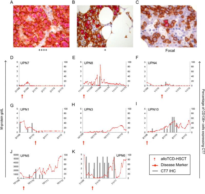
Figure 1. CT7 is expressed in CD138+ plasma cells in the BM of myeloma patients and is associated with the disease course.

CT7 protein expression in CD138+ plasma cells was monitored longitudinally over the course of the disease in MM patients undergoing alloTCD-HSCT. Paraffin-fixed BM biopsies from MM patients were double stained with monoclonal antibodies to CD138 (MI15; DAB, brown) and CT7 (CT7-33; nFu, red). Immunohistochemical analysis of CT7 expression was performed and biopsies were graded negative, focal, +, ++, +++ or ++++ based on the percentage of CD138+ PC staining positive for CT7. Representative biopsy stainings are shown in Figures A–C. (A) ++++ >75% of CD138+ plasma cells stained positive for CT7; UPN6 (B) + >5–25% of CD138+ cells are CT7+; UPN5. (C) Focal, <5% of CD138+ cells are CT7+; UPN1 (20×). Longitudinal analyses of CT7 protein expression presented in panels D-K. The percentages of CD138+ cells expressing CT7 were compared to each patient’s relevant disease marker (D-I/K, M-Spike gamma g/dl; J, IgA mg/dl). (D) UPN7 (E) UPN8 (F) UPN4 (G) UPN1 (H) UPN3 (I) UPN10 (J) UPN5 (K) UPN6.
