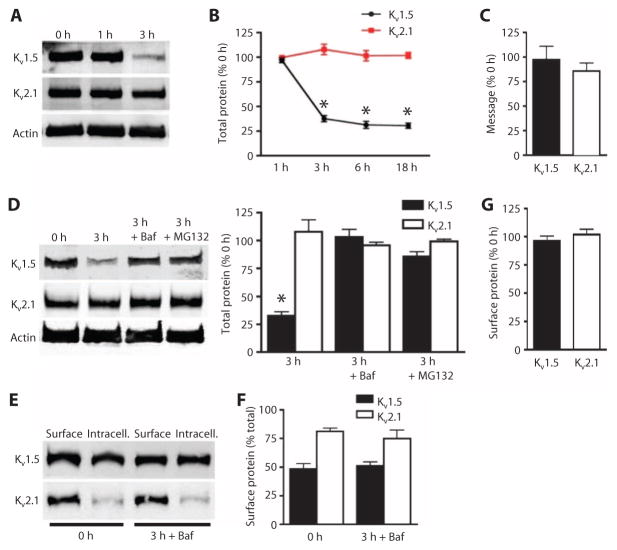
Fig. 2. Bafilomycin and MG132 prevent Kv1.5 loss in mesenteric arteries.
(A) Representative Western blot images of total Kv1.5 and Kv2.1at 0, 1, and 3 hours of isolation. (B) Quantification of total Kv1.5 and Kv2.1 at the indicated times after isolation. n = 5 for the 1-hour group, n = 6 for the remaining groups. *P < 0.05 versus 0 hour. (C) Quantitative PCR data expressed as the percent of transcripts for each channel remaining after 3 hours with the amount of transcript at 0 hour set at 100%. n = 3 for each. (D) Representative Western blots and quantification of total Kv1.5 and Kv2.1 0 and 3 hours after arterial isolation, and the effect of bafilomycin (Baf; 50 nM) or MG132 (10 μM). n = 6 for each. *P < 0.05 versus 0 hour. (E) Representative Western blot images of arterial biotinylation samples showing the abundance of surface and intracellular Kv1.5 and Kv2.1 at 0 and after 3 hours of arterial isolation in the presence of bafilomycin. (F) Mean data showing the percent of total Kv1.5 and Kv2.1 at the cell surface for arteries immediately and 3 hours after isolation. n = 5 for each. (G) Mean data showing percent of Kv1.5 and Kv2.1 remaining at the cell surface after 3 hours in the presence of bafilomycin relative to the 0-hour samples. n = 5 for each.

