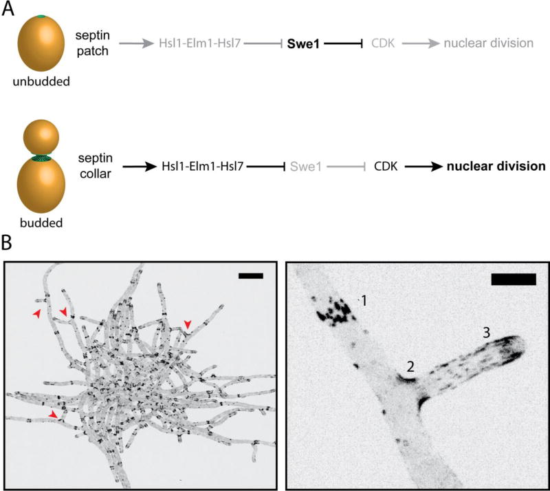
Figure 3. Septin structures and localization in budding yeast and the filamentous fungus Ashbya gossypii.
A. Morphogenesis checkpoint in S. cerevisiae: Hsl1-Elm1-Hsl7 are not recruited to the septin patch in unbudded cells, thereby allowing the CDK-inhibitor Swe1 levels to rise. As soon as the bud emerges, septin localization transitions to an hourglass shape coincident with the generation of micron-scale curvature. Swe1-regulators are recruited after this transition to initiate the degradation of Swe1 and promote progression through the cell cycle.
B. An inverted, maximum z-stack projection of a A. gossypii cell expressing Cdc11-GFP. Scale bar, 20 µm. (Bottom right) Zoomed-in image of A. gossypii hyphae expressing Cdc11-GFP from above. Septins are organized into various higher-ordered structures in Ashbya: [1] Thick bars structures, [2] basal collars at branch points, and [3] thin, flexible filaments. Scale bar, 5 µm.

