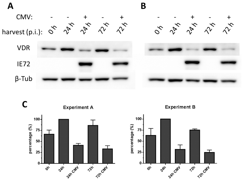
Figure 3. Downregulation of VDR protein generation during early and late phases of CMV infection.
HFF were infected with CMV AD169 (MOI 3) or mock-infected and treated with (A) calcitriol at a final concentration of 1 nM or (B) calcitriol (1 nM) together with the VDR agonist EB-1089 at a final concentration of 100 nM. Cell lysates were probed with VDR-specific antibody to test for VDR protein expression (VDR), an anti-IE72 antibody to verify presence of virus in the infected samples (IE72) and an antibody specific for beta-tubulin to confirmed equal protein amounts loaded on the blotting membrane (β-Tub). C: From two experiments, each bands’ signal intensity was normalized to the signal intensity of the corresponding loading control (β-Tub). Percentage of each bands’ signal intensity considering the strongest band as 100 % was calculated and mean values and SEM (n=2) of the percentage evaluation are shown.

