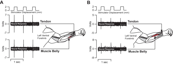
Fig 3. Bypassing the myotendinous junction.
(A&B) Two representative single unit peripheral nerve recording traces from the median nerve with schematic drawings of the limb tendons and musculature. The neural recordings were maintained while the stimulator pulled on the freed tendon with a 2 mm displacement. The upper neural recording trace shows responses through three cycles with the stimulator pulling on the freed distal tendon. The lower trace shows three cycles of the same afferent after the stimulator was moved to the muscle belly to bypass the myotendinous junction. The nerve signal was maintained in each position.

