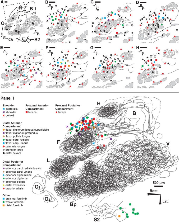
Fig 4. Cortical brain mapping of the RA-MS response group elicited by tapping the tendons of the degloved forelimb.
Symbols represent individual electrode penetrations: X = No Response; Red Stars = Cutaneous responses before paw denervation; Black Stars = Cutaneous responses after paw denervation. Scale bars = 500 μm; R = rostral; L = lateral. (A) Overview of the left cortical hemisphere in the rat. Cytochrome oxidase delineated borders and barrel patterns of primary somatosensory cortex (S1) (black lines and grey shading): F = forepaw barrel subfield; H = hindpaw barrel subfield; V = vibrissae; Bp = buccal pad; L = lower lip, O1 & O2 = oral modules 1 & 2; L = chin/lower lip; B = body; S2 = second somatosensory area. The dashed box denotes the area of interest enlarged in B through H. (B-H) The RA-MS response group projected to a region of cortex anterior and adjacent to the forepaw barrel subfield and the area between the caudal lower lip and forelimb representations. Recordings at each electrode penetration typically yielded responses from only a single muscle of the forelimb. The RA-MS responses were entirely exclusive from the cytochrome oxidase defined borders of the somatotopic S1 cutaneous representation. (I) A scaled composite overlay of all seven recording cases showing the global organization of the muscle-specific tapping-sensitive RA-MS responses in the rat. Although RA muscle afferents did project to areas near the forepaw and caudal lower lip, the majority of afferents projected to the darkly staining region connecting the forelimb and hindlimb representations (dashed lines). The random organization of the RA-MS unit responses is evident. In contrast, single electrode penetrations in the second somatosensory area (S2) had receptive fields that encompassed the entire forelimb.

