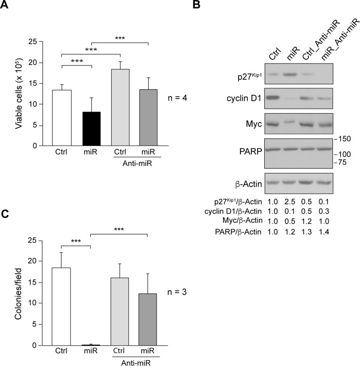
Fig 2. Role of miR205 in cell proliferation and anchorage-independent growth.
(A) Cell proliferation was determined by performing Trypan blue exclusion assay in SUM159_Control (Ctrl), SUM159_miR205 (miR), SUM159_Control_Anti-miR205 (Ctrl_Anti-miR), SUM159_miR205_Anti-miR205 (miR_Anti-miR) cells. Cells were seeded at 3x10⁵ cells/60mm dishes, and 72h later both Trypan blue-negative (viable cells) and Trypan blue-positive cells (dead cells) were counted (see Materials and Methods). Results are shown as measurements of Trypan blue-negative cells, mean ± SD from four independent experiments in triplicate. (B) Immunoblotting detection of p27Kip1, cyclin D1, Myc, and PARP in cell extracts. Membranes were reblotted with anti-β-actin for loading control. Results are representative of three independent experiments. The ratios referred to SUM159_Control (Ctrl) considered as 1. (C) Number of colonies/field obtained after cell growth in soft-agar for 10 days (see Material and Methods). Average of colony number/field ± SD from three independent experiments in triplicate. (***p<0.001).

