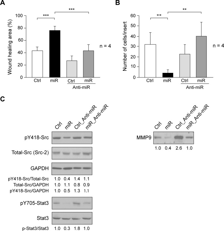
Fig 3. Effect of miR205 expression on cell migration and invasion.
(A) Cell migration was determined by wound-healing assay in SUM159_Control (Ctrl), SUM159_miR205 (miR), SUM159_Control_Anti-miR205 (Ctrl_Anti-miR), SUM159_miR205_Anti-miR205 (miR_Anti-miR) cells (see Materials and methods). Results are expressed as mean percentage of wound healing area ± SD at 20h respect to 0h from three independent experiments (***p < 0.001). (B) Cell invasion through MatrigelTM-coated inserts (see Materials and methods). The number of invaded cells per insert is shown as average ± SD of four experiments in triplicate. (**p<0.01). (C) Immunoblotting detection of activated SFKs (pY418-Src), Total-Src (Src2), and GAPDH as loading control, as well as pY705-Stat3, and Stat3 in cell extracts, and of MMP9 in cellular secretome from SUM159_miR205 and Sum159_Control cells containing equal amount of total proteins. They are representative results from 3 independent experiments carried out in triplicate. The ratios referred to SUM159_Control (Ctrl) considered as 1.

