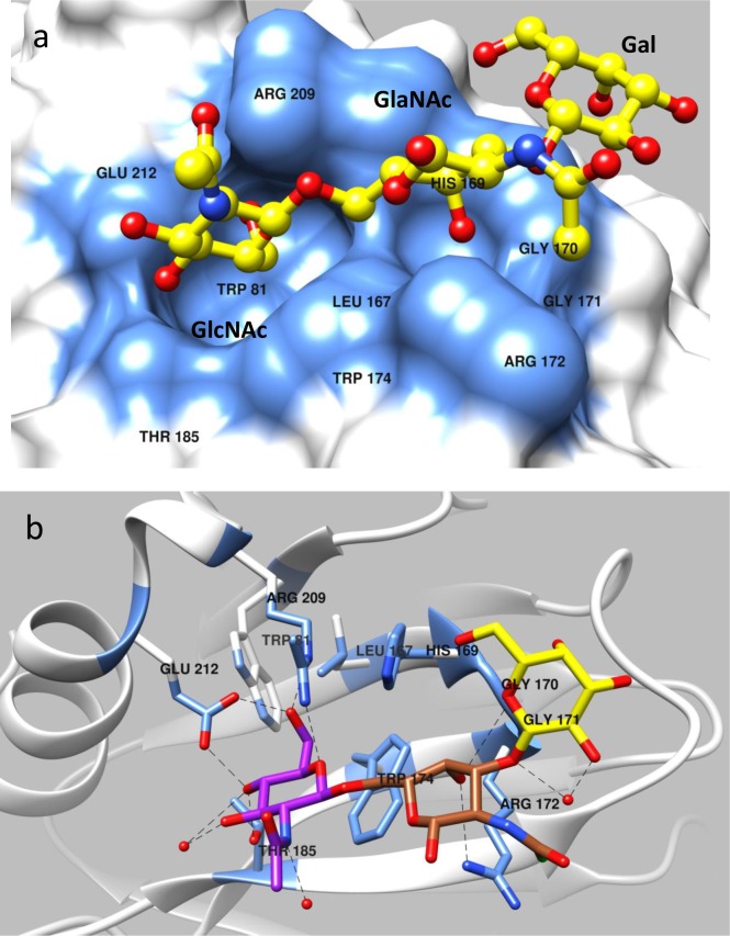
Fig 4. Structure of P[19] VP8* in complex with mucin core 2.
(a) Surface representation of P[19] VP8* structure with bound mucin core 2 following the same coloring scheme as in Fig 2a. (b) Network of hydrogen bond interactions between the VP8* residues and mucin core 2 with the same coloring scheme as in Fig 2b. All the three sugar moieties of mucin core 2 are involved in interaction with the VP8*, with the GlcNAc and GalNAc of mucin core 2 (GlcNAcβ1-6GalNAcβ1-3Gal) binding to the two adjacent well-defined pocket on the surface of P[19] VP8*.

