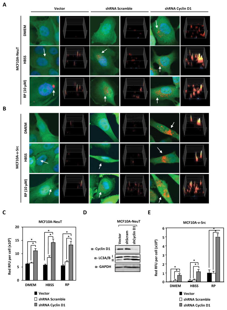
Figure 2. Endogenous cyclin D1 restrains stress-induced autophagy in oncogene transformed human breast cancer cells.
(A) MCF10A-NeuT and (B) MCF10A-v-Src cells expressing RFP-LC3 were treated with shRNA against cyclin D1 or control shRNA in the presence of either DMEM, HBSS or Rapamycin (RP) (10 μM) for 2 hours. Immunofluorescence detection of RFP-LC3 shows increased LC3 redistribution to autophagosomes (yellow/red perinuclear dots) in cells transduced with shCCND1. (C) Quantitation of LC3 puncta/cell area in MCF10A-NeuT and (D) Western blotting on the same cells demonstrates increased LC3-II in MCF10A-NeuT. (E) Quantification of LC3 puncta/cell area in MCF10A-c-Src (expressed in square pixels, n=50). Cyclin D1 reduces autophagosome formation (* P<0.05 and ** P<0.01).

