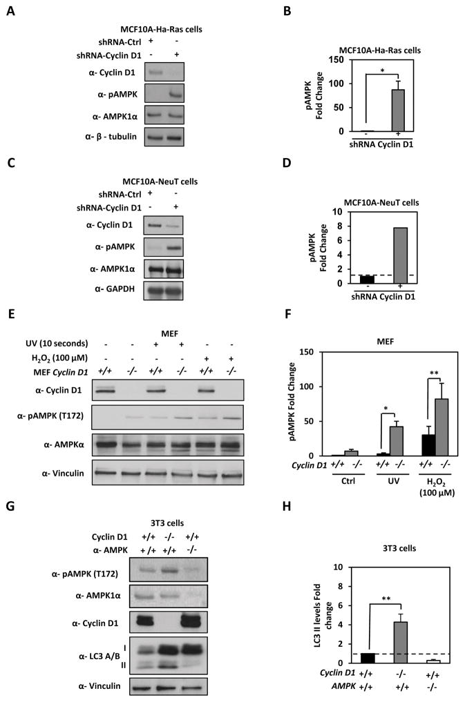
Figure 3. Endogenous cyclin D1 restrains stress induced AMPK.
(A) MCF10A-Ha-Ras and (C) MCF10A-NeuT expressing cyclin D1 shRNA were analyzed by Western blotting for LC3A/B-II, AMPK and pAMPK. Quantitation of pAMPK levels in (B) MCF10A-Ha-Ras and (D) MCF10A-NeuT, fold enrichment of pAMPK is shown. (E) cyclin D1+/+ and cyclin D1−/− MEF were treated with cellular stressors (UV and H2O2) and cell lysates analyzed by Western blot as shown. (F) Fold enrichment of pAMPK were quantified and shown. (G) cyclin D1+/+ and cyclin D1−/− 3T3 cells compared to Ampk1α2α−/− 3T3 by Western blot to analyze phosphorylated Ampk (T172), lipidated LC3-II levels, cyclin D1 and normalized to vinculin to calculate LC3-II levels (H)(* P<0.05; ** P<0.01 and *** P<0.001).

