. 2017 Nov 28;6:e27364. doi: 10.7554/eLife.27364

Figure 2. Rational optimization of tetrapeptide ACC substrates with iP and cP selectivity.

(A) Example 14-mer peptides from the iP and cP MSP-MS assays associated with Figure 1 illustrating cleavages that were used as a template for rational fluorogenic substrate design. All cleavages common between two biological replicates of each proteasome are reported with the time point of first appearance noted for each replicate. (B) Selectivity of parental IQ and tetrapeptide ACC fluorogenic substrates derived from an iP-favored cleavage (Figure 2A, top) in the MSP-MS library. Subsequent rational substrate optimization is shown. (C) Michaelis-Menten characterization and chemical structure of the lead iP substrate, EWFW-ACC. (D) Optimization of an ACC substrate with cP selectivity using a similar approach. Shortening the peptide sequence to a tripeptide and addition of an N-terminal capping group were found to be important for improving substrate selectivity and maintaining specific activity. (E) Michaelis-Menten characterization and chemical structure of the lead cP substrate iso-VQA-ACC. For all fluorogenic substrate assays, mean activity is reported with error bars representing the standard deviation from n = 3 replicates.

Figure 2.

Figure 2—figure supplement 1. P4-P2 substrate specificity of SDS-activated iP and cP using the PS-SCL profiling approach.

Figure 2—figure supplement 1.

Mean activity is reported with error bars representing the standard deviation from n = 3 replicates.
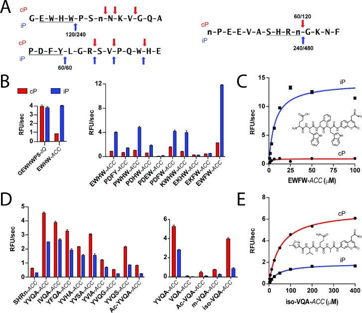
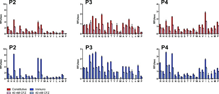
Figure 2—figure supplement 2. PS-SCL profiling of SDS-activated iP and cP in the absence and presence of 40 nM CFZ pretreatment.

Figure 2—figure supplement 2.

CFZ levels were selected based on inhibitor titration against the library to achieve optimal selectivity. Mean activity is reported with error bars representing the standard deviation from n = 3 replicates.
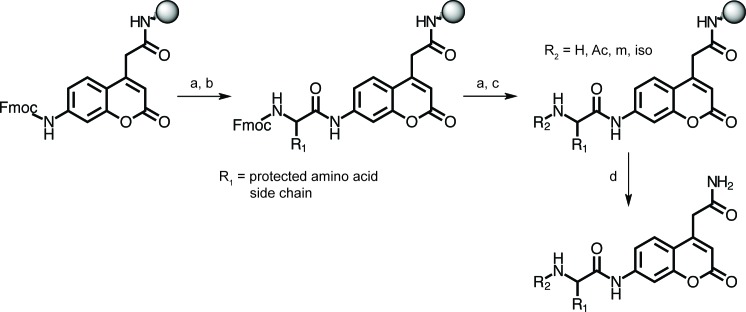
Figure 2—figure supplement 3. Synthetic approach for peptide substrates bearing a C-terminal ACC fluorophore.

Figure 2—figure supplement 3.

Protocols for synthesis of the Fmoc-ACC fluorophore and its coupling to Rink amide resin have been described. (Maly et al., 2002) (a) 20% 4-methylpiperidine/DMF; (b) Fmoc-protected P1 amino acid (5 eq.), HATU (5 eq.), and 2,4,6-collidine (5 eq.), DMF, 24 hr, or Fmoc-protected P2, P3, or P4 amino acid (6.5 eq.), HBTU (6.5 eq.), and N-methylmorpholine (13 eq.), DMF, 1 hr; (c) for acetyl (Ac): acetic anhydride/TEA/DMF/DCM (1:1:5:5), for N-morpholinyl (m): 4-morpholinylacetic acid hydrochloride (6.5 eq.), HBTU (6.5 eq.), and N-methylmorpholine (26 eq.), and for 5-methylisoxazolyl (iso): 5-methylisoxazole-3-carboxylic acid (6.5 eq.), HBTU (6.5 eq.), and N-methylmorpholine (13 eq.); (d) TFA/H2O/triisopropylsilane (95:2.5:2.5).
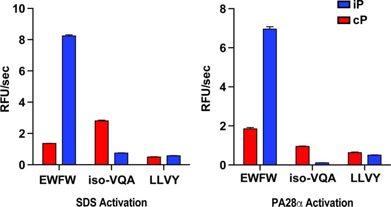
Figure 2—figure supplement 4. Comparison of fluorogenic substrate selectivity under SDS (0.03%) and PA28 (12 eq.) activation conditions.

Figure 2—figure supplement 4.

Mean activity is reported with error bars representing the standard deviation from n = 3 replicates.