
Figure 2. Physical interaction between HEXIM1 and AR.
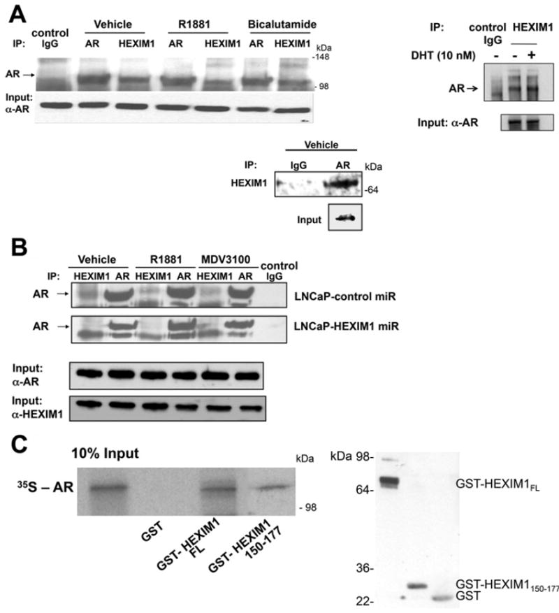
(A) LNCaP cells were treated with either vehicle, 10 nM R1881, 10 nM DHT (dihydrotestosterone) or 10 μM bicalutamide for 90 min. (B) LNCaP cells stably transfected with control or HEXIM1miR were treated with vehicle, 10 nm R1881 or 10 μM MDV3100 for 90 min. In (A) and (B) lysates were immunoprecipitated using antibodies against HEXIM1 or AR and analysed for co-immunoprecipitation of HEXIM1 or AR by Western blotting. Normal rabbit IgG was used as a specificity control. Input lanes represent 25% of the total protein. (C) Left-hand panel: in vitro translated and [35S]methionine-labelled AR was incubated with GST alone, GST–HEXIM1 or GST–HEXIM1150–177 bound to Sepharose. Bound protein was eluted and analysed by SDS/PAGE (12.5% gel). The Input lane represents 10% in vitro translated product added to the samples. Right-hand panel, Western blot analyses of GST, GST–HEXIM1 or GST–HEXIM1150–177 expression. Figures are representative of at least three independent experiments.