Figure 4.
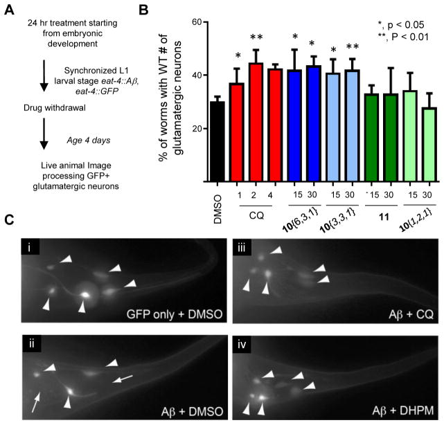
DHPM-thiones rescue nematode model of Aβ toxicity. (A) Schematic of the compound treatment regimen for nematode Aβ model. (B) Percent of worms with wild-type number of glutamatergic neurons labeled with eat-4::GFP. Three independent experiments were performed for each compound (concentration tested in μM) where 30 worms were scored in each trial (for a total of 90 worms). P-Values comparing DMSO to each compound were calculated using a one-way ANOVA and Tukey’s test. (C) Representative fluorescent images of GFP-labeled tail-region glutamatergic neurons in the Aβ nematode model for GFP only (i), Aβ plus DMSO (ii), Aβ plus CQ (iii), and Aβ plus compound 10{6,3,1} (iv). Large arrowheads indicate neurons. Small arrows with a line indicate a missing neuron.

