
Fig. 3.
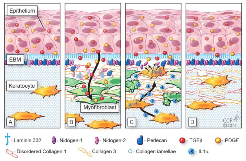
Schematic diagram illustrating injury to the corneal epithelial basement membrane (EBM) and defective regeneration leading to myofibroblast development and fibrosis, followed by hypothesized keratocyte contributions to regeneration of the EBM and resolution of fibrosis. A) Normal unwounded cornea with intact epithelial basement membrane (EBM) comprised of laminin 332, nidogen-1, nidogen-2, perlecan, and other components not depicted such as collagen type IV. The underlying stroma is populated with fibroblastic cells (keratocytes) that function to maintain the highly-organized stromal collagen lamellae that provide the cornea transparency. Epithelial transforming growth factor beta (TGFβ) and platelet-derived growth factor (PDGF) are blocked from penetration into the underlying stroma by the normal EBM. B) After severe epithelial-stromal injuries, such as infections, trauma and some surgeries, the epithelium and EBM are disrupted and TGFβ and PDGF are activated and penetrate the underlying stroma at sufficient concentrations to drive the development of myofibroblasts from keratocyte-derived and bone marrow-derived (fibrocyte) precursors [16]. Myofibroblasts are themselves opaque relative to keratocytes and secrete disordered collagen type 1, collagen type 3 and other matrix materials that disrupt the normal stromal lamellae to produce corneal opacity or scarring. C) Over months to years following the initial injury, keratocytes penetrate the anterior stromal myofibroblasts and facilitate EBM regeneration via the production of laminins, nidogens, and perlecan [38,39,43] in coordination with epithelial cell production of EBM components. We hypothesize that once the nascent laminin-332 layer is produced by the epithelium, more posterior EBM components must, at least in part, be derived from keratocytes to fully regenerate the EBM. The resulting decrease in TGFβ and PDGF penetration from the epithelium into the stroma triggers myofibroblast apoptosis via unopposed paracrine IL-1α from adjacent keratocytes and/or autocrine IL-1α produced by the myofibroblasts themselves (autocrine suicide) [17]. This process begins in a random spotty distribution within the stromal opacity to produce clear areas of stroma called “lacunae” (Fig. 1C) that enlarge and coalesce over weeks to months to fully restore transparency. D. In many corneas, depending on the severity of the injury, all myofibroblasts disappear and keratocytes fully-repopulate the stroma and reabsorb the remaining disorganized collagens and matrix materials secreted by the myofibroblasts to completely restore the normal morphology of the collagen lamellae and, thereby, stromal transparency.