
Figure 1. Cultured cells overexpressing wild type human α-synuclein accumulate oligomeric aggregates of α-synuclein.
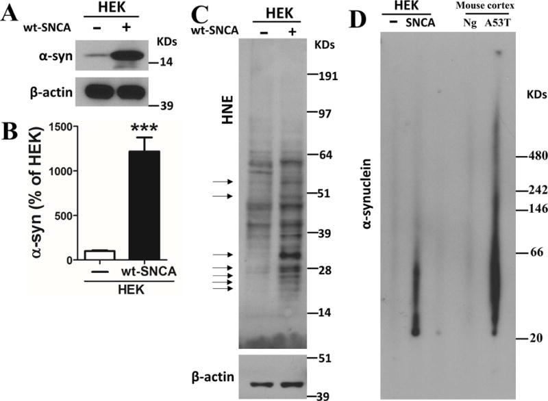
(A) Immunoblot showing relative levels of α-synuclein and β-actin as an internal control in HEK cells with or without human wildtype α-synuclein (wt-SNCA) overexpression. (B) Results of densitometric analysis of α-synuclein levels in HEK cells with or without human wildtype α-synuclein overexpression. Values are the mean and SEM of determinations made in 9 independent experiments. ***p<0.001. (C) Immunoblot showing relative levels of HNE-protein adducts and β-actin in HEK cells with or without human wild type α-synuclein overexpression. Arrows point to protein bands that exhibit much higher levels of 4-hydroxynonenal (HNE) immunoreactivity in samples from the cells overexpressing wild type α-synuclein. (D) Lysates prepared from HEK cells with or without α-synuclein overexpression, and cerebral cortex from nontransgenic (Ng) and Thy1-A53T-SNCA transgenic mice were run in NativePAGE™ Bis-Tris gels. The immunoblot reveals high amounts of α-synulcein aggregates in samples of HEK cells overexpressing α-synuclein and cortical tissue from Thy1-A53T-SNCA transgenic mice, compared to respective control HEK cells and wild type mice.