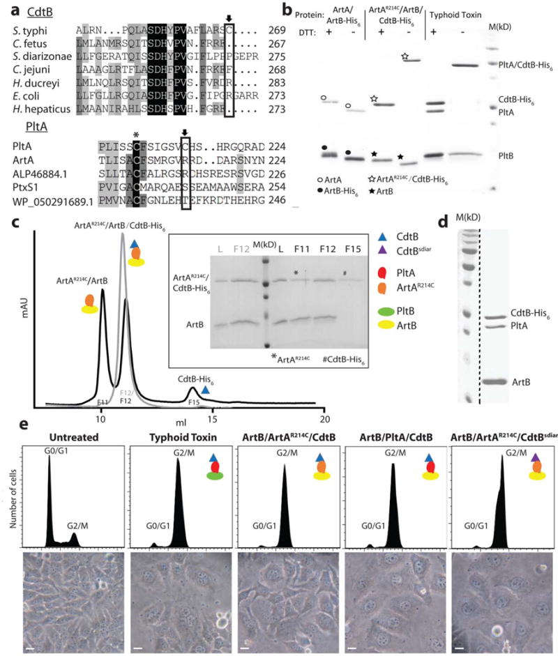
Figure 1.

The ArtAB toxin components can form a functional complex with typhoid toxin subunits. a, Amino acid sequence comparison of CdtB and PltA homologs. Conserved and unique cysteines are indicated with an asterisk and an arrow, respectively. ALP46884.1 and WP_050291689.1 are PltA homologs from Escherichia coli and Yersinia kristensenii, respectively. PltxS1 is the A subunit from pertussis toxin. b, Purified ArtB/ArtA, ArtB/ArtAR214C/CdtB, or typhoid toxin (PltB/PltA/CdtB) protein complexes were analyzed by SDS-PAGE in the presence or absence of DTT to release CdtB, which is linked to the complex by a disulfide bond. The migration of ArtA and His6-tagged CdtB are very similar in SDS-PAGE so in the presence of DTT the two bands overlap. In the absence of DTT, ArtA and CdtB (like CdtB and PltA in the case of typhoid toxin, see right lanes) migrate as a single, slower moving band, an indication that these subunits are linked by a disulfide bond. ○: indicates ArtA; ●: indicates ArtB-His6; ☆: indicates ArtAR214C/CdtB-His6; ★: indicates ArtB. This experiment was carried out twice with equivalent results. c, The ArtAR214C/ArtB/CdtB chimeric toxin complex was analyzed by ion exchange chromatography before (gray) and after (black) treatment with DTT. L: loading control; M: molecular weight markers; F: chromatographic fraction. Inset shows SDS-PAGE analyzes of the indicated chromatographic fractions. *: ArtAR214C; #: CdtB-His6. This experiment was carried out once. d, ArtB can form a complex with wild-type PltA and CdtB. The ArtB/PltA/CdtB complex was purified by ion exchange and size exclusion chromatography and subsequently analyzed by SDS-PAGE and coomassie blue staining. This experiment was carried out three times with equivalent results. The dashed black line indicates that this panel is a composite image of two discontinuous lanes from the same gel. e, Toxicity of the chimeric toxin complexes. Cultured Henle-407 epithelial cells were treated with Typhoid Toxin (3.5 pM), ArtB/ArtAR214A/CdtB (15 pM), ArtB/PltA/CdtB (15 pM), or ArtB/ArtAR214A/CdtBsdiar, and the CdtB-mediated cell cycle arrest was assayed by flow cytometric analysis. CdtBsdiar: CdtB from S. diarizonae. Light microscopic images of mock or toxin treated cells are also shown. Scale bar: 50μm. This experiment was carried out three times with equivalent results.
