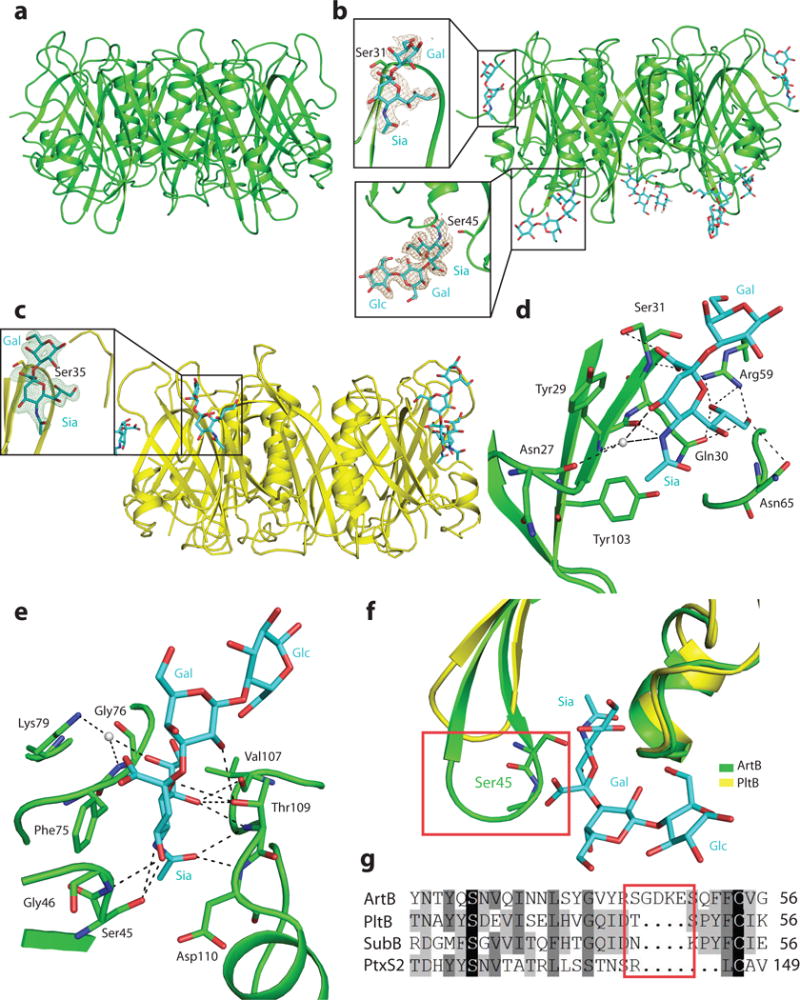
Figure. 3.

The atomic structure of ArtB bound to its receptor shows the presence of an additional glycan-binding site. a, Atomic structure of the ArtB pentamer shown as a ribbon cartoon. b, Atomic structure of the ArtB pentamer in complex with the Neu5Acα2–3Galβ1–4Glc oligosaccharide shown as a ribbon cartoon. Cyan, blue and red sticks represent carbon, nitrogen and oxygen atoms in the sugar backbone. Insets show the close-up views of Neu5Acα2–3Galβ1–4Glc and Neu5Acα2–3Gal. Brown mesh represents the sugar composite annealed omit difference density map contoured at 2.0σ. c, Atomic structure of the PltB pentamer in complex with the Neu5Acα2–3Gal oligosaccharide is shown as a ribbon cartoon. Cyan, blue and red sticks represent carbon, nitrogen and oxygen atoms in the sugar backbone. Insets show the close-up views of Neu5Acα2–3Galβ1–4Glc. Green mesh represents the sugar composite annealed omit difference density map contoured at 2.5σ. d and e, Interactions between ArtBSer31 (d) and ArtBSer45 (e) with Neu5Acα2–3Gal and Neu5Acα2–3Galβ1–4Glc, respectively. ArtB is shown as a green colored ribbon cartoon, the sugar and the amino acids interacting with the sugar are shown as sticks, the interactions are shown in black dashes and water is shown as gray balls. f, Structural comparison of ArtBSer45 sugar-binding site with the equivalent surface in PltB. Blue and red sticks in the sugar backbone represent nitrogen and oxygen atoms, respectively. g, Amino acid sequence alignment of ArtBSer45 glycan-binding site with the equivalent regions in PltB, SubB and PtxS2. The red boxes depicted in f and g highlight the insert sequence and the associated structural features that are uniquely present in ArtB.
