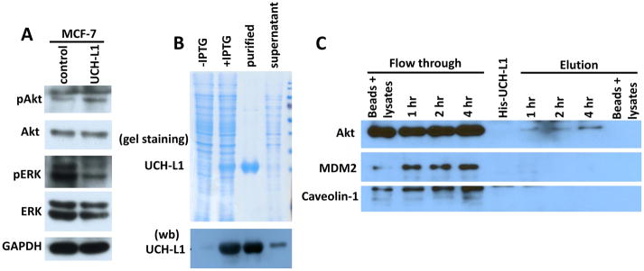
Figure 3. UCH-L1 activates Akt signaling pathway.
(A) MCF-7 cells were transfected with plasmid to generate stable UCH-L1 overexpression cell line. Overexpression of UCH-L1 led to activation of Akt as evidenced by upregulation of p-Akt level. (B) A His tagged recombinant human UCH-L1 expression plasmid was generated and transformed into E. Coli. Coomassie blue staining of SDS-PAGE (upper panel of B) and Western blotting (lower panel of B) show that His-rhUCH-L1 was successfully purified by Ni-NTA column. (C) His-rhUCH-L1 was used as a bait to pulldown interacting proteins from cell lysates derived from MCF-7 cells. Western blot analysis show that Akt can be pulled down by His-rhUCH-L1 protein, while other proteins, such as MDM2 and Cavin-3 cannot be found in elution fraction of the experiments. Empty Ni-NTA beads incubated with cell lysates and purified His-rhUCH-L1 were also included as controls. n = 3 independent experiments.

