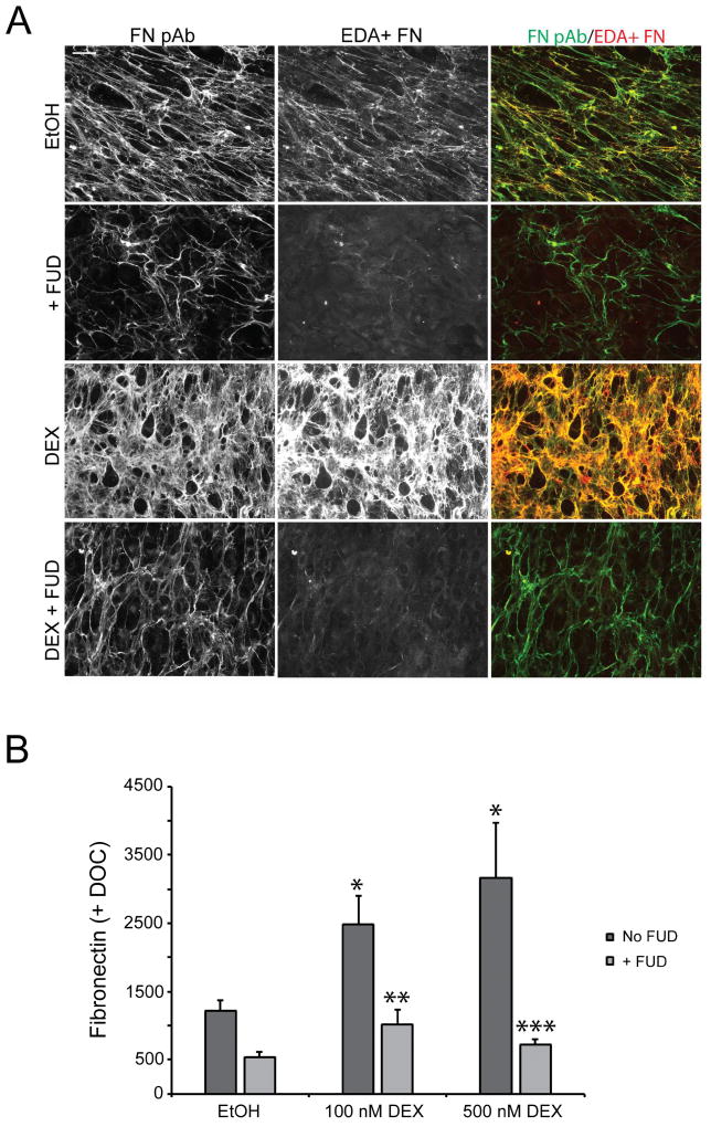
Figure 4. FUD blocks steroid-induced increases in fibronectin fibrillogenesis and matrix assembly.
A) Confluent cell monolayers were treated with vehicle (EtOH) or 500 nM DEX for 12–14 days in the presence or absence of 2 µM FUD prior to fixation and double labeling with fibronectin antisera (FN pAb) and mAb IST-9 against EDA+ FN. B) Cell monolayers in 96 wells plates were treated as in panel A. We used the fibronectin polyclonal antisera in this assay rather than mAb IST-9 as the antisera would detect the multiple isoforms of fibronectin present in our HTM cultures as suggested by the immunofluorescence microscopy results described in panel A. At the end of the treatment period, cell monolayers were extracted with 1% DOC prior to processing for OCW analysis. *, significantly greater than EtOH-treated cells, p < 0.01; **, significantly less than DEX (100nM) treated cells without FUD, p < 0.05; ***, significantly less than DEX (500nM) treated cells without FUD, p < 0.01. Scale bars = 50 µm.

