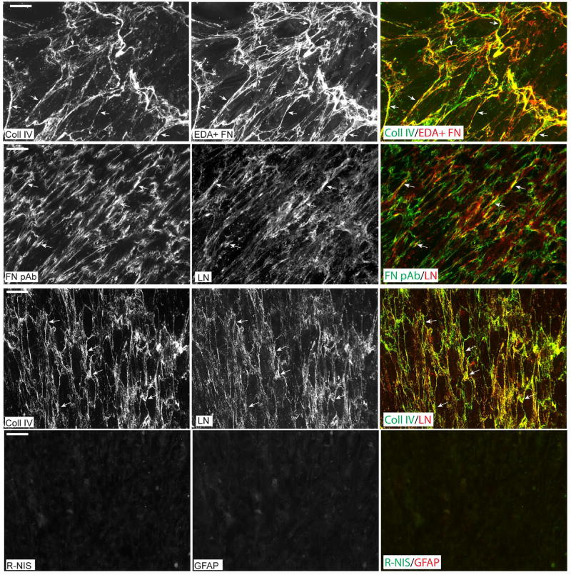
Figure 5. Co-localization of ECM proteins expressed by HTM cells.
HTM cells that had been maintained at confluence for 7 days were double-labeled for type IV collagen and EDA+ fibronectin (top row), fibronectin and laminin (2nd row) or type IV collagen and laminin (3rd row). Arrows indicate areas of co-localization for the different antibody pairs. Cultures were also double-labeled with rabbit non-immune serum and a mAb against glial fibrillary acidic protein (GFAP) which served as controls for antibody specificity (4th row). Cultures in row 1 were methanol-fixed while the other three rows show labeling in paraformaldehyde-fixed cells. Negative control labeling was also performed in methanol-fixed cells and showed similar results (not shown).

