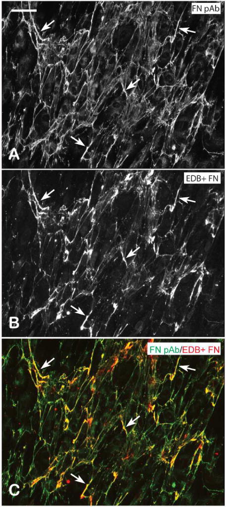
Figure 9. Fibronectin fibrils consist of a mixture of EDB+ and EDB- fibronectin isoforms.
Confluent HTM cells were fixed and labeled with fibronectin polyclonal antisera (A) and mAb BC-1 against EDB+ fibronectin (B). The merged images (C) show select regions of co-localization of the two antibodies (arrowheads). Scale bar = 50 um.

