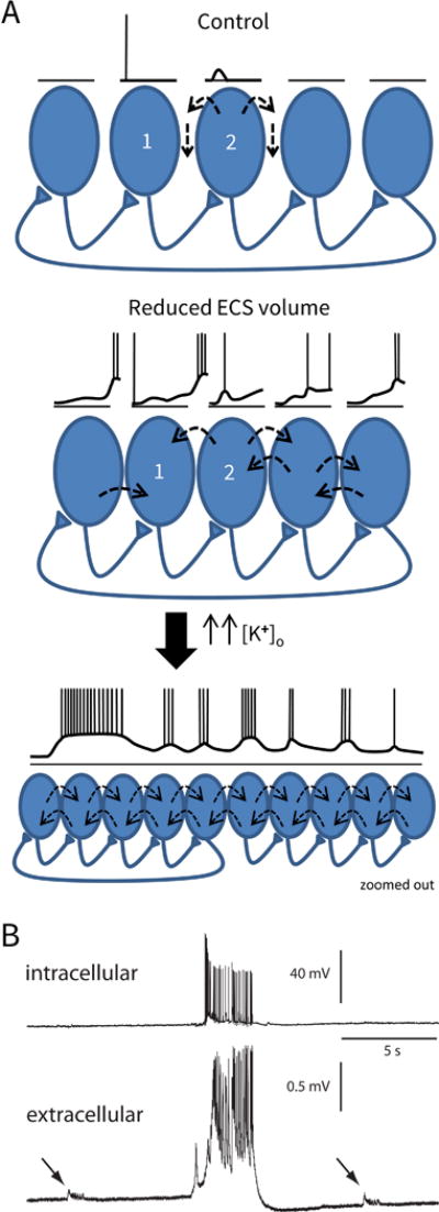
Figure 5.

Generation of epileptic activity in the condition of reduced ECS volume.
(A) Model. Top panel is the control situation. CA1 pyramidal cell 1 fires an action potential which causes an epsp in the postsynaptic cell (2), but cell 2 does not fire. Current leaving cell 2 takes the paths of least resistance, and thus mostly travels in the ECS, and not across the cell membranes of the neighboring cells. (Dashed arrows depict current flow.) In the middle panel, the ECS volume is reduced (as in Has3−/− mice). There is a small increase in [K+]o because the K+ which leaves firing cells is diluted less in the smaller ECS volume. This small increase in [K+]o causes a small baseline depolarization of the cells, and now when cell 1 fires, the epsp in cell 2 triggers an action potential. Because the cells are closer together and extracellular resistance is larger, some of the current leaving cell 2 travels across the membranes of neighboring cells, causing them to depolarize (ephaptic interactions). Epsps in cells further down the chain overlap with depolarizations caused by ephaptic interactions, producing larger responses. (The longest voltage sequence is shown for cell 1.) Interictal activity will begin to occur as the depolarizations synchronize among the cells, which will cause a further rise in [K+]o. In the lower panel, a larger population of cells has been recruited. Both the rise in [K+]o and a fall in [Ca+]o will increase cell excitability and enhance the persistent Na+ current, which, in this condition of enhanced ephaptic interactions, will enable the recruitment of a larger population and allow ictal-like events to occur. (The view is zoomed out. The drawn voltage trace applies for all of the cells.) (B) Actual voltage traces recorded in the CA1 region of a Has3−/− brain slice. Top trace is an intracellular recording from a single pyramidal cell. Bottom trace is a simultaneous recording of the population field potential. The tiny interictal-like events (arrows) are occurring in a population of cells that does not include the neuron from which the intracellular recording is taken. However, the large, ictal-like event recruits a larger population of neurons, including the one from which the intracellular recording is taken. (B is modified from Fig. 4C in Arranz et al., 2014).
