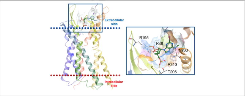
FIGURE 3.

X-ray structure of the hP2Y1R showing the binding mode of orthosteric antagonist MRS2500 (3) [38]. In this inactive state, the 3′-phosphate is coordinated by K46 (EL1) and R195 (EL2), the 5′-phosphate deeper in the binding site by T205 (EL2) and R310 (7.39) and N6 by N283 (6.58).
