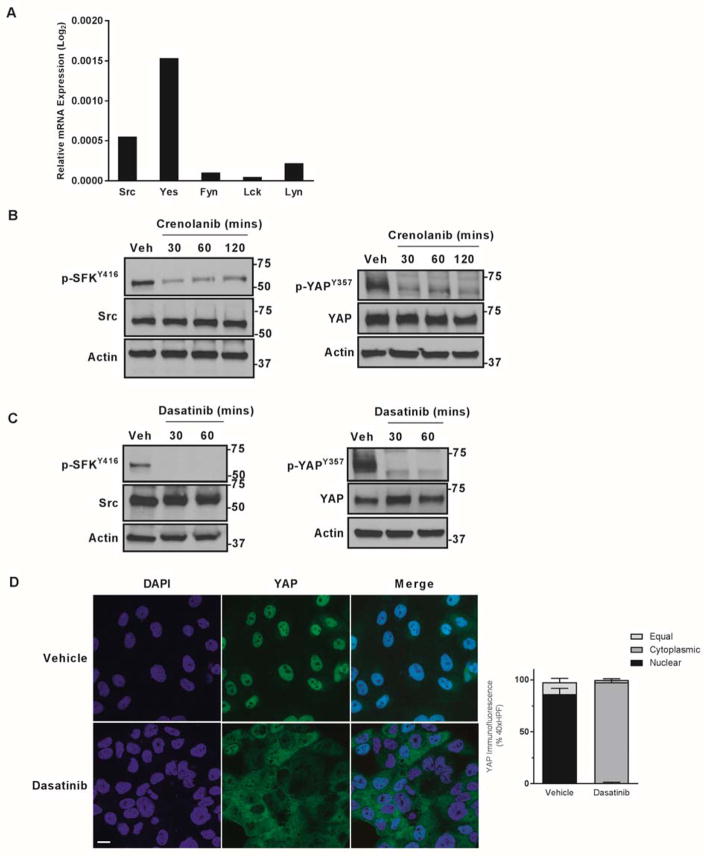
Figure 4. PDGF mediated YAP tyrosine phosphorylation is via Src Family Kinases.
A, mRNA expression in HuCCT-1 cell lines. Relative expression compared to 18S internal control is depicted as geometric mean (n=3). B,C cell lysates from the HuCCT-1 cell line ± PDGFR inhibitor crenolanib (10 μM) (B) or the SFK inhibitor dasatinib (1 μM) (C) were subjected to immunoblot for phosphorylated Src (Y416) and Src and phosphorylated YAP (Y357) and YAP. Actin was used as a loading control. D, immunofluorescence images from the HuCCT-1 CCA cell line ± SFK inhibitor dasatinib (1 μM) with quantification of subcellular localization (right panel). Scale bar = 10 microns.

