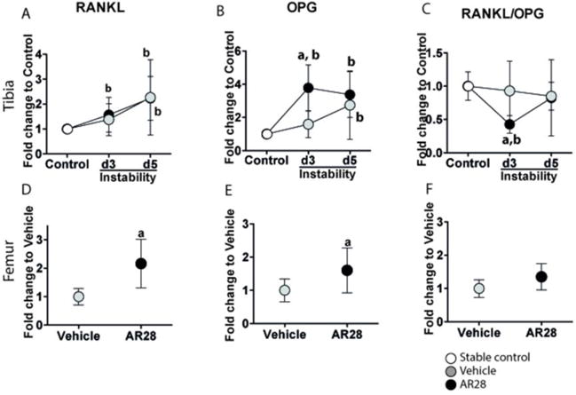
Figure 6.

GSK-3β inhibition suppresses osteoclast differentiation in an OPG-associated manner in the bone underneath the implant. Relative mRNA expression of RANKL (A, D), OPG (B, E) and RANKL/OPG ratio (C, F) in the tibial implant bone (upper panels) and intact femoral bone (lower panels). Results are presented as the fold-change in mRNA values compared to the non-loaded controls (0 days) in tibial bone and to vehicle-treated animals in femoral bone. Graphs indicate mean±SD in each group. a: significant compared to vehicle-treated animals, b: significant changes compared to controls. A p-value of <0.05 was considered significant.
