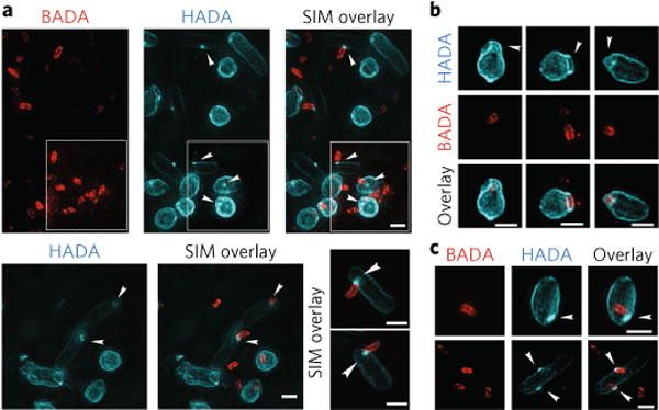
Fig. 2. Three-dimensional structured illumination microscopy images of early predation by B. bacteriovorus (pre-labelled with BADA, false-coloured red) on prey E. coli cells after pulse labelling for 10 min with HADA (false-coloured cyan) to show early modification of cell walls.

a, Predation 15min post-mixing reveals a ring of HADA-labelled prey cell wall modification at the point of B. bacteriovorus contact (arrowheads) and of similar width to the B. bacteriovorus cell (see Supplementary Table 2). Central pores in the labelled PG material can be seen where the B. bacteriovorus image is artificially removed from the overlay of the two channels. Such annuli may represent a thickened ring of PG modification. In the white inset, the lookup table for the BADA channel has been separately adjusted until all the BADA labelled predators are clearly visible. Three representative examples are displayed. b, Prey PG is deformed around the site of B. bacteriovorus invasion (arrowheads). c, The cells show HADA fluorescence at the end of the internal B. bacteriovorus cell (arrowheads), which probably represents transpeptidase activity re-sealing the hole in the prey PG after the B. bacteriovorus cell has entered. Images are representative of >100 3D-reconstructed cells in two independent experiments (Supplementary Table 2 for details of numbers analysed). Scale bars, 1 μm.
