
Fig. 3. Three-dimensional structured illumination microscopy images of early predation by B. bacteriovorus (pre-labelled with BADA, false-coloured green) on prey E. coli imp4213 cells (which are more permeable and thus susceptible to the TADA pre-labelling, false coloured in red) after pulse labelling for 10min with HADA (false-coloured cyan) to show early modification of cell walls.
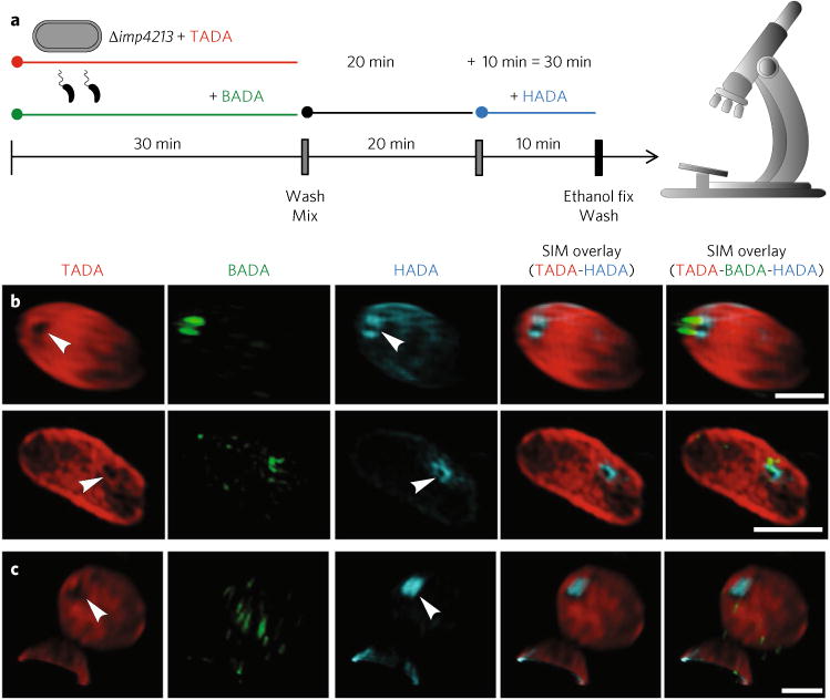
a, FDAA labelling scheme (using excess B. bacteriovorus to promote synchronous invasion of E. coli Δimp4213 mutant prey) with time points observed by 3D-SIM fluorescence microscopy. Predator and prey cells were pre-labelled separately with BADA and TADA, respectively, before being washed and then mixed. Samples of this mixed infection were then pulse-labelled with HADA for 10min before the time points of 15 or 30min. The cells were then fixed, washed and microscopically observed. b, Predation 30 min post-mixing with this prey strain reveals a pore in the TADA signal coincident with the ring of HADA-labelled prey cell wall modification at the point of B. bacteriovorus contact (arrowheads) and of similar width to the B. bacteriovorus cell (Supplementary Table 3). c, In several cases (Supplementary Table 3) where the B. bacteriovorus cell had entered into the prey cell and established itself in the periplasm of the bdelloplast, the pore in the TADA was coincident with a patch of HADA labelling and thus is likely to represent the sealing of the pore through which the B. bacteriovorus had entered. Images are representative of two independent experimental repeats. Scale bars, 1 μm.