Figure 1.
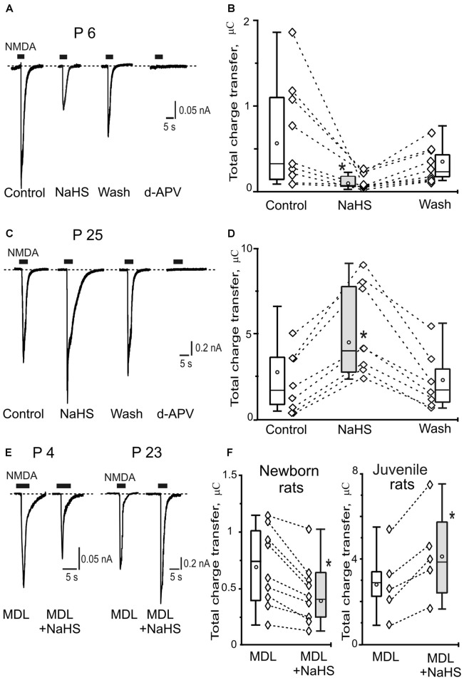
Age-dependent effects of sodium hydrosulfide (NaHS) on N-methyl-D-aspartate (NMDA) evoked currents in CA3 area of rat hippocampus. Representative current traces recorded from pyramidal neurons at a holding potential −60 mV activated by application of 100 μM NMDA + 30 μM glycine (5 s, black bars) in control, in the presence of 100 μM NaHS, after washout and after inhibition of NMDA receptors by d-2-Amino-5-phosphopentanoate (dAPV; 40 μM) at the age P6 (A) and at the age P25 (C). Statistical plot of NMDA induced total charge transfer in control, in the presence of NaHS and after washout in pyramidal neurons of neonatal (B; n = 9, N = 6) and juvenile rats (D; n = 8, N = 6). Each pair of connected diamonds corresponds to an individual neuron. The mean values of boxplots are shown by white circles, whiskers—minimal and maximal values. (E) Representative current traces recorded from pyramidal neurons activated by application of 100 μM NMDA + 30 μM glycine (5 s, black bars) after incubation with the inhibitor of adenylate cyclase MDL-12330A (MDL; 10 μM) and in the presence of MDL+NaHS at the age P4 and P23. (F) Statistical plot of NMDA induced total charge transfer in the presence of MDL and MDL+NaHS in pyramidal neurons of neonatal (n = 9; N = 6) and juvenile (n = 5; N = 4) rats. Each pair of connected circles corresponds to individual neurons. Boxes indicate 25–75 percentiles in control (white) and in NaHS (gray), black line—median, the circle inside—mean value, whiskers—minimal and maximal values, *p < 0.05, paired W-test.

