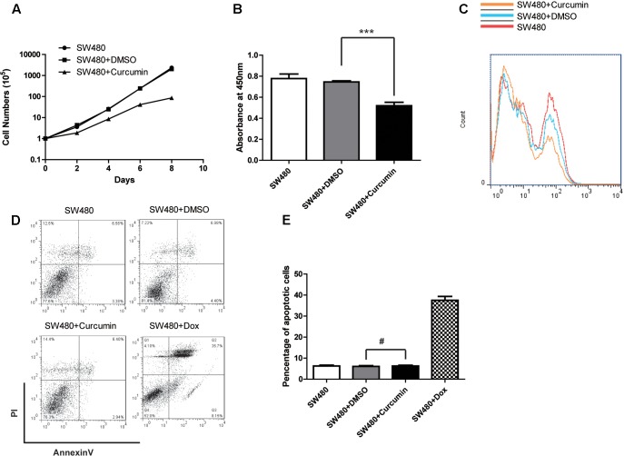
FIGURE 2.
Curcumin inhibited the cell proliferation of SW480. (A) Cell proliferation of different groups as indicated was determined by cell counting. The concentration of curcumin was 40 μM. Data were represented as mean ± SD; n = 3 independent experiments. (B) CCK-8 kit was used to evaluate the viability of each group of the cells as indicated. Data were represented as mean ± SD; n = 3 independent experiments. ∗∗∗p < 0.001. (C) Cell proliferation was detected by BrdU incorporation assay and n = 3 independent experiments and this panel presented one of these repeats. (D) The apoptosis of the SW480 cells with different treatment as indicated was determined by using the Annexin-V-FITC & PI Apoptosis Kit and assessed by flow cytometry. n = 3 independent experiments and this panel presented one of these repeats. (E) Statistic of percentage of the apoptosis cells performed in (D). Data showed the Annexin-V and PI double positive cells. Data were represented as mean ± SD; n = 3 independent experiments. #p > 0.05.

