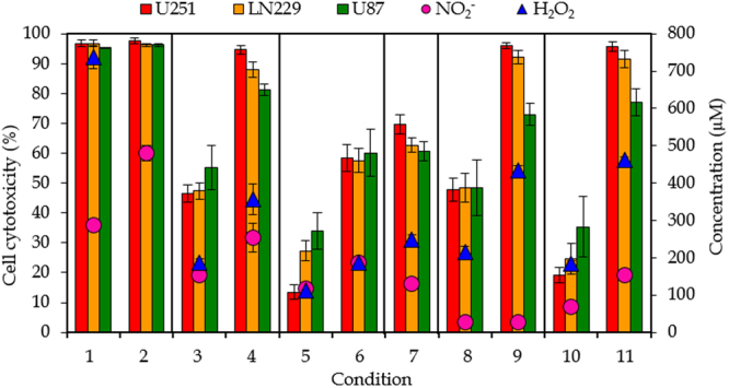
Figure 4.

General overview of the results. Effect of pPBS on cancer cell cytotoxicity for three different GBM cell lines (U251, LN229 and U87) (left y-axis), and comparison with the concentrations of NO2 − and H2O2 in pPBS (right y-axis), for the 11 conditions listed in Table 1. Note that the H2O2 concentration in condition 2 is 1317 µM, but this is deliberately out of scale, to better evaluate the correlation between cell cytotoxicity and chemical composition for all other conditions. Conditions that only differ in plasma treatment time are indicated within one frame. The concentrations and percentages are plotted as the mean of at least three repetitions, and the error bars indicate the standard deviations of the mean.
