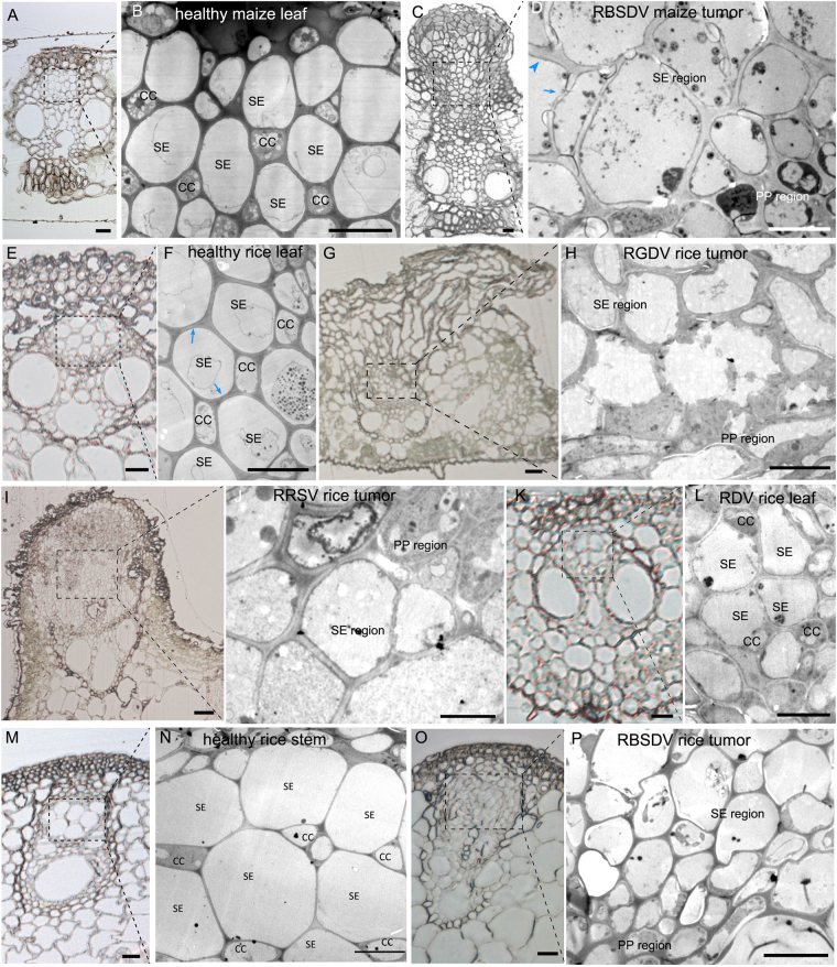
Figure 2.
Re-organization of cells in plant-reoviral infected phloem. (A) Histological section of healthy maize leaf vascular bundle. (Bar = 20 μm). (B) Magnification of healthy maize leaf phloem in (A) under transmission electron microscopy (TEM) showing its cellular arrangement with a staggered SE-CC pattern. (Bar = 10 μm). (C) Histological section showing the cellular hyperplasia of the phloem in the infected vascular bundle causing the tissue to erupt through the epidermis as a tumor. (Bar = 20 μm). (D) Magnification of RBSDV-infected maize phloem in (C) under TEM showing how SEs are aggregated into exclusive regions without CC. (Bar = 10 μm). Most SEs in the tumor have thick CWs but thickening was not homogeneous (thickened part marked by blue arrowhead and the thin part by blue arrow). (E) Histological section showing the healthy vascular bundle of rice leaf. (Bar = 10 μm). (F) Magnification of healthy phloem of rice leaf in (E) under TEM showing its cellular arrangement with staggered SE-CC pattern. (Bar = 5 μm). (G) Histological section showing the cellular hyperplasia of the phloem in the infected vascular bundle which erupted through the epidermis as a tumor. (Bar = 20 μm). (H) Magnification of RGDV-infected rice phloem in (G) under TEM showing how SEs are aggregated into exclusive regions without CC. (Bar = 10 μm). (I) Histological section showing the cellular hyperplasia of the phloem in the infected vascular bundle which erupted from the epidermis as a swollen vein. (Bar = 20 μm). (J) Magnification of RRSV-infected rice phloem in (I) under TEM showing how SEs are aggregated into exclusive regions without CC. (Bar = 5 μm). (K) Histological section showing the non-hyperplasia of RDV-infected rice phloem. (Bar = 10 μm). (L) Magnification of RDV-infected rice phloem in (K) under TEM showing its cellular arrangement with staggered SE-CC. (Bar = 5 μm). (M) Histological section of healthy vascular bundle without hyperplasia. (Bar = 20 μm). (N) Magnification of healthy phloem in (M) under TEM showing its cellular arrangement with a staggered SE-CC pattern. (Bar = 10 μm). (O) Histological section showing the cellular hyperplasia of the phloem in the infected vascular bundle but without eruption into a tumor. (Bar = 20 μm). (P) Magnification of RBSDV-infected rice phloem in (O) under TEM showing how SEs are aggregated into exclusive regions without CC. (Bar = 10 μm).

