
Figure 1.
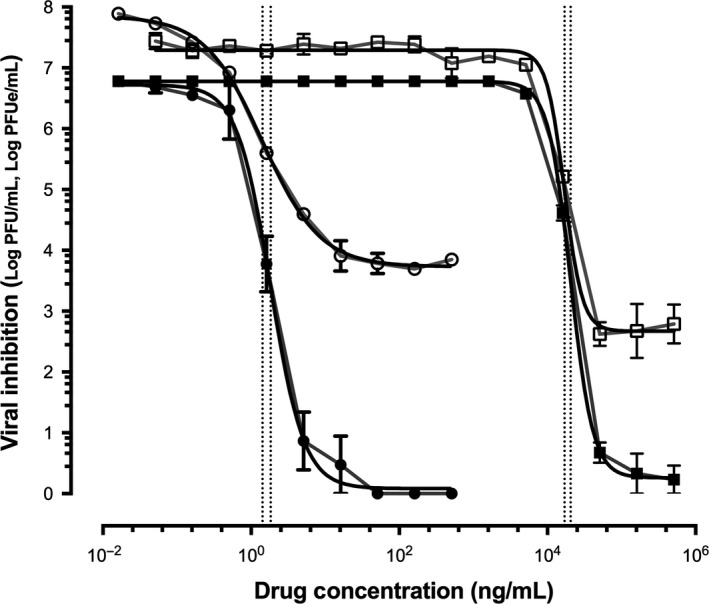
Inhibition of Respiratory syncytial virus (RSV)‐A Long by MDT‐637 (represented by closed and open circles) and ribavirin (represented by closed and open squares) measured by quantitative culture (represented by closed circles and closed squares, quantification unit; log plaque forming units/mL, log PFU/mL) and quantitative PCR (represented by open circles and open squares, quantification unit; log plaque forming unit equivalents/mL, log PFUe/mL). The vertical dotted lines represent the IC 50 for MDT‐637 (1.83 ng/mL by quantitative culture and 1.42 ng/mL by quantitative PCR) and ribavirin (20 509 ng/mL by quantitative culture and 16 973 ng/mL by quantitative PCR)