Figure 1.
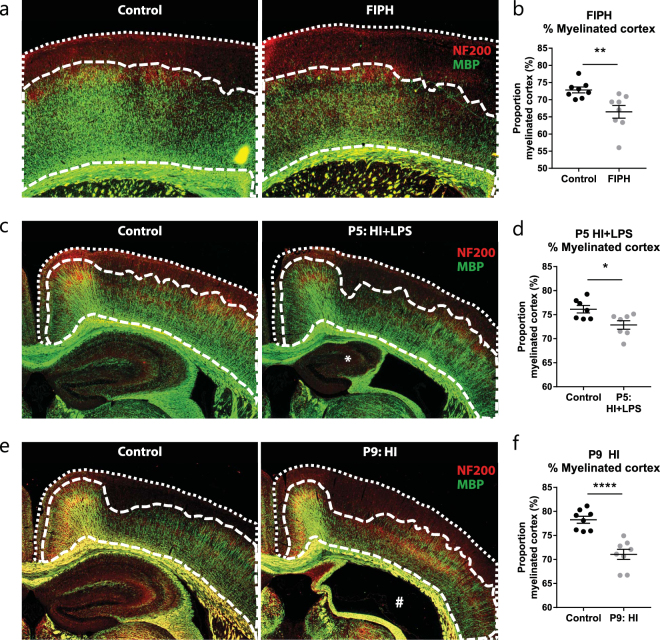
Neonatal brain injury is associated with reduced myelination in outer cortical layers. (a) Representative micrographs of a control rat (left panel) and a rat exposed to fetal inflammation (LPS at E18 + E19) and postnatal hypoxia at P4 (FIPH model; right panel) stained for axonal marker NF200 (red) and myelin marker MBP (green). (b) Bar graph showing that rats exposed to FIPH display reduced extent of cortical myelination (n = 8 per group). (c) Representative images of sham-operated control mice (left) or mice exposed to HI + LPS at P5 (right) stained for MBP (green) and NF200 (red). *Enlarged ventricle and reduced ipsilateral hippocampal volume which is typically observed in this model. (d) Bar graph illustrating that HI + LPS mice show reduced extent of cortical myelination (n = 6 per group). (e) Representative images of sham-operated control mice (left) or mice exposed to HI on P9 (right) stained for MBP (green) and NF200 (red). #: severely injured hippocampal area characteristic for this model (f) Bar graph illustrating that HI mice show reduced extent of cortical myelination (n = 8 per group). Data represent mean ± SEM; *p < 0.05; **p < 0.01; ****p < 0.0001.

