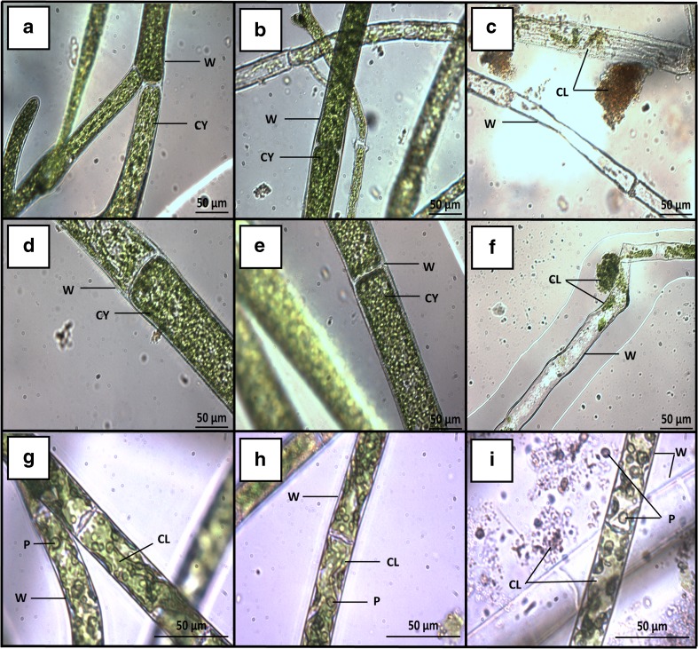
Fig. 4.
Plates of C. coelothrix (a–c), C. parriaudii (d–f), and S. varians (g–i) taken with an inverted microscope after a 14-day growth trial (100 rpm, 24 °C, light intensity of 30–40 μmol photons m−2 s−1, 18:6 h L/D photoperiod) with frequent harvesting using different methods described in Table 1: positive control (+ C) (a, d, and g), beaker + reticulated spinner (B+RS) (b, e), beaker + filter paper (B+FP) (c, f, and i), and perforated crucible + reticulated spinner (PC+RS) (h). W denotes the cell wall, CL indicates the chloroplasts, P is the pyrenoid, and CY highlights the multi-nucleate cytoplasm that contains pyrenoids, chloroplasts, and vacuoles

