Figure 6.

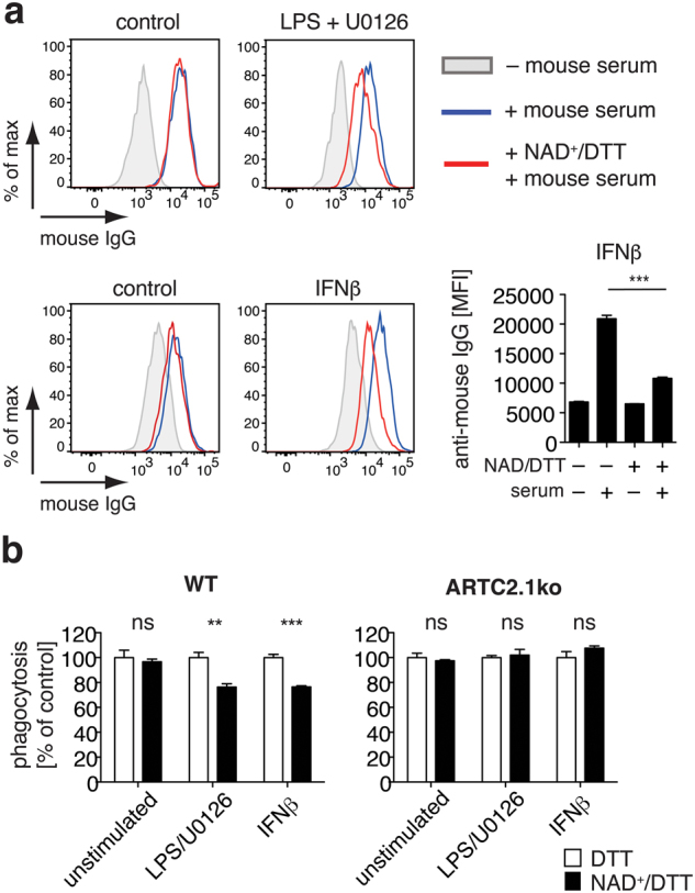
ADP-ribosylation of FcγRs affects binding of IgG to microglia. (a) IgG binding to unstimulated, LPS/U0126-stimulated (upper plot) or IFNβ-stimulated (lower plot) microglia was analyzed by flow cytometry. Cells were pre-incubated with NAD+/DTT or left untreated, washed twice with PBS and were incubated with IgG containing mouse serum (1:20 diluted) for 30 min. Cells were washed twice and cell surface binding of mouse IgG was measured using a PE-conjugated anti-mouse IgG F(ab)2. (b) Phagocytosis of PE-labeled IgG-coated latex beads by unstimulated, LPS-stimulated or IFNβ-stimulated microglia from WT or ARTC2.1−/− mice was measured by flow cytometry. Cells were either preincubated with DTT alone or with NAD+/DTT for 30min, washed twice and allowed to phagocytose IgG-beads for 3h. Phagocytosis rate was normalized to the corresponding DTT control sample (100%). Data are representative of 2–3 independent experiments. Statistical comparison of two groups was performed by using the student’s t test (p < 0.05 = */p < 0.01 = **/p < 0.001 = ***).
