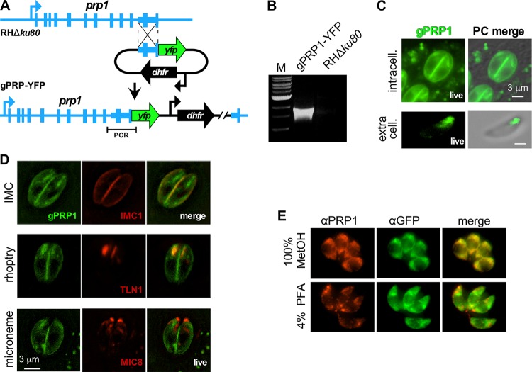
FIG 4 .
Subcellular localization of PRP1 through endogenous tagging. (A) Schematic representation of generating C-terminal endogenously YFP-tagged gPRP1-YFP parasites by single homologous recombination into the RHΔku80 parent line. (B) PCR validation of the gPRP1-YFP genotype using the primer pair shown in panel A. Lane M contains 1-kb DNA ladder (New England Biolabs). (C) Live imaging of gPRP1-YFP parasites under intracellular and extracellular conditions as indicated. PC, phase contrast. (D) Live imaging of gPRP1-YFP parasites cotransfected with markers for the IMC (IMC1-mCherry), rhoptries (TLN1-mCherry), and micronemes (MIC8-mCherry). (E) Representative images of intracellular gPRP1-YFP parasites fixed using either 100% methanol (MetOH) or 4% paraformaldehyde (PFA) stained with anti-PRP1 (αPRP1) and anti-GFP (αGFP) antisera as indicated. Note that PFA fixation destroys the costaining of GFP and PRP1 and thus destroys the PRP1 epitope(s) recognized by the specific antiserum.

